What Are Cherry Angiomas?
Cherry angiomas, also known as Campbell de Morgan spots or senile angiomas, are small, benign (noncancerous) growths made up of clustered small blood vessels (capillaries). They appear as bright red or purplish spots on the skin, often resembling a mole but with a distinctly vascular nature.
Common Characteristics
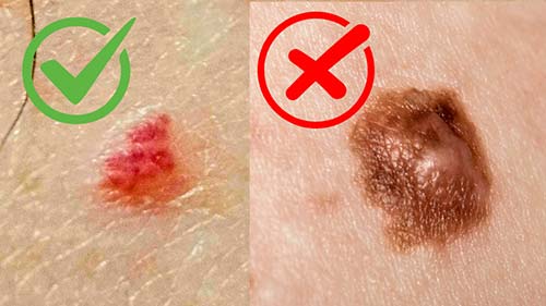
- Appearance: Bright cherry-red to purple, round or oval, flat or slightly raised dome-shaped; may have a pale halo around them3
- Size: Typically 1-5 mm in diameter, though they can grow larger over time
- Texture: Smooth and soft; may bleed easily if scratched, traumatized, or caught on clothing
- Locations: Most common on the trunk (chest, back, abdomen), arms, legs, and neck; rarely on hands, feet, face, or mucous membranes1
- Symptoms: Usually none — no pain, itching, or discomfort unless irritated; purely cosmetic concern for most people
Are Cherry Angiomas Dangerous?
Cherry angiomas are entirely benign and have no relationship to cancer whatsoever. They are not contagious and pose no health risks. However, if many appear suddenly (eruptive cherry angiomas) or if one changes significantly in size, shape, or color, medical evaluation is recommended to rule out underlying conditions like immunosuppression or lymphoproliferative disorders.1
How Cherry Angiomas Form — The Science
Cherry angiomas develop from an overgrowth and clustering of capillaries in the skin's dermis layer. This creates a small, dilated vascular network that pushes toward the surface as a red spot. The vessels within become tortuous (twisted) and dilated, creating the characteristic dome-shaped appearance.
Recent genetic research has identified somatic mutations in GNAQ and GNA11 genes in about 50% of cherry angiomas tested.4 These same mutations appear in other vascular lesions like port-wine stains and certain hemangiomas, suggesting a shared genetic pathway. Additionally, reduced levels of microRNA-424 have been found in cherry angiomas, leading to elevated growth factors (MEK1) that promote abnormal blood vessel formation.5
What Causes Cherry Angiomas?
The exact cause of cherry angiomas isn't fully understood, but research has identified several contributing factors. They become significantly more common with age and often run in families, suggesting both genetic and environmental components.
Age & Genetics
The primary risk factors. Cherry angiomas rarely appear before age 30 but increase dramatically with advancing age — from about 5% prevalence in young adults to over 75% in those over 75.1 Strong genetic predisposition means if close relatives have many cherry angiomas, you're significantly more likely to develop them.
Genetic Mutations
Research has identified specific somatic mutations in GNAQ and GNA11 genes in about 50% of cherry angiomas.4 These mutations affect cellular signaling pathways that control blood vessel growth, explaining why some people develop numerous angiomas while others develop few or none.
Hormonal Changes
More common during pregnancy due to increased blood volume, vascular changes, and hormonal shifts. Some pregnancy-related cherry angiomas resolve spontaneously postpartum, but many persist.6
Sun Exposure
Cumulative UV damage may contribute, as they often (but not exclusively) appear on sun-exposed areas. However, cherry angiomas also commonly develop on covered areas like the trunk, suggesting sun is not a primary cause but may be a contributing factor.
Chemical Exposure
Rare associations with exposure to certain chemicals like topical nitrogen mustard (used for vitiligo treatment), bromides, and butoxyethanol have been documented.7 Eruptive cherry angiomas can occur as a side effect of these exposures.
Other Associations
Possible links to certain medical conditions including immunosuppression, graft-versus-host disease, lymphoproliferative disorders, and multi-centric Castleman disease. Some studies suggest associations with lipid abnormalities, though evidence is limited.8
When to See a Doctor: If cherry angiomas multiply rapidly (eruptive cherry angiomas), bleed without cause, change appearance significantly, or are accompanied by other concerning symptoms, consult a healthcare provider. While harmless themselves, sudden onset may indicate underlying conditions worth investigating.
⚠️ Critical First Step: Verify It's Actually a Cherry Angioma
Before attempting ANY treatment method (home or professional), proper identification is essential. What appears to be a cherry angioma could be something else requiring different care or medical attention.
Common Look-Alikes
- Petechiae or Purpura - Flat red spots from bleeding under skin; don't blanch with pressure
- Pyogenic Granuloma - Similar vascular growth but often larger, bleeds very easily, trauma-related
- Spider Angiomas - Star-shaped with visible central vessel; linked to liver disease or hormones
- Amelanotic Melanoma - Can appear red/pink; irregular, grows rapidly, serious concern
- Angiokeratomas - Dark red to black; warty texture; different structure
Warning Signs - Seek Medical Evaluation
- Asymmetrical shape or very irregular borders
- Multiple colors within the growth (not just red/purple)
- Spontaneous bleeding without trauma
- Rapid growth over weeks rather than gradual over years
- Pain or tenderness without obvious irritation
- Hard texture or feels attached to deeper tissue
Our Professional Assessment Process
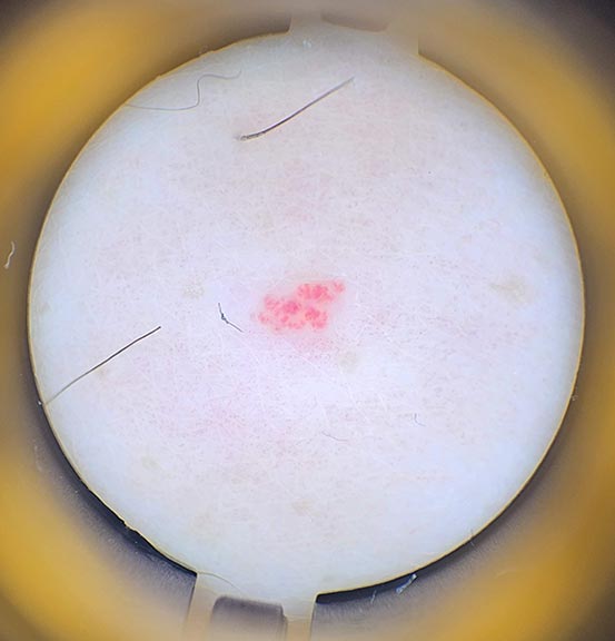
At Hideaway Spa, every growth is examined using a dermatoscope (dermlite) before treatment. This specialized magnification tool allows detailed visualization of vascular structures to:
- Confirm the growth is a benign cherry angioma by visualizing dilated capillary loops
- Rule out concerning features like irregular vascular patterns suggesting malignancy
- Assess vascular density and depth for appropriate treatment planning
- Document baseline appearance for clinical records
Any lesion exhibiting atypical vascular patterns, suspicious features, or clinical uncertainty is referred to a dermatologist before treatment proceeds.
⚠️ Never treat a growth without being absolutely certain of what it is. Cherry angiomas have characteristic features, but only a trained professional can distinguish them from potentially serious conditions.
Cherry Angioma Treatment Options: Complete Comparison
Once confirmed as benign, cherry angiomas don't require treatment unless for cosmetic reasons or if they bleed frequently. Several evidence-based methods exist, each with distinct advantages and limitations.
| Method |
How It Works |
Best For |
Scarring Risk |
Time to Result |
Cost (Approx.) |
| Pulsed Dye Laser (PDL) |
Laser targets hemoglobin in blood vessels; heat destroys vessels which fade over time |
Multiple small angiomas; preferred by many dermatologists for minimal pain |
Low-Moderate |
1-2 weeks (darkens first) |
$200-500+ |
| Nd:YAG Laser |
Deeper penetrating laser coagulates blood vessels |
Darker skin tones (less pigmentation risk than PDL) |
Low-Moderate |
1-2 weeks |
$200-500+ |
| Cryotherapy |
Liquid nitrogen freezes tissue (-196°C); vessels destroyed by ice crystal formation |
Small to medium angiomas; widely available |
Moderate |
10-14 days |
$100-300 |
| Shave Excision |
Surgical blade shaves off growth flush with skin surface |
Large, raised angiomas; when biopsy needed |
Moderate-High |
Immediate (7-10 day healing) |
$150-400 |
| Thermolysis/Electrocautery (Our Method) |
Radiofrequency probe instantly coagulates blood vessels; angioma visibly clears during treatment |
Any size; multiple angiomas; all skin tones; instant results desired |
Very Low |
Instant visible clearance (full healing 7-14 days) |
$50-200 |
Cost estimates reflect typical Windsor/Ontario market rates as of 2026. Individual needs and outcomes vary.
Understanding Risks & Complications for All Methods
Every treatment method carries potential risks. Understanding these helps you make informed decisions and set realistic expectations.
Pulsed Dye Laser (PDL) - Risks
- Purpura (bruising): Expected side effect lasting 7-14 days; purple discoloration before fading
- Pigmentation changes: Hyper/hypopigmentation possible, especially in darker skin tones (Fitzpatrick IV-VI)
- Incomplete clearance: May require 2-3 sessions for complete removal
- Blistering: Rare but possible with aggressive settings
- Cost: Most expensive option; multiple sessions increase total cost significantly
Nd:YAG Laser - Risks
- Less pigmentation risk: Safer for darker skin than PDL but still carries some risk2
- Deeper penetration: Can cause more discomfort during treatment than PDL
- Multiple sessions: Often requires 2-3 treatments for full clearance
- Cost: Similar to PDL; expensive option
Cryotherapy - Risks
- Hypopigmentation: Permanent skin lightening common, especially in darker skin tones
- Blistering and pain: Significant blistering for 7-14 days; can be uncomfortable
- Infection risk: Blisters can become infected without proper care
- Incomplete treatment: May need multiple freeze cycles; less reliable than other methods
- Higher recurrence: 15-20% recurrence rate — highest among professional methods9
Shave Excision - Risks
- Bleeding: Immediate bleeding requiring cauterization or pressure
- Scarring: Higher scarring risk; may leave depressed or raised scar
- Infection: Any surgical wound carries infection risk
- Anesthesia required: Local injection needed (brief pinch/burn)
- Longer healing: 2-4 weeks for complete healing vs days for other methods
Thermolysis/Electrocautery (Our Method) - Risks
- Temporary pigmentation: Mild hyper/hypopigmentation possible but typically resolves within weeks
- Small crust formation: Expected healing response lasting 3-7 days (tiny scab)
- Temporary redness: Treatment site may remain slightly pink for 7-14 days
- Infection (rare): Any skin injury carries infection risk with improper aftercare
- Low recurrence: 5-10% recurrence rate — among lowest of all methods9
- Scarring (minimal): Lowest scarring risk of all methods when properly performed10
Proper aftercare significantly reduces all risk factors. Strict sun avoidance and no picking at crusts essential.
All risk information based on dermatological literature and clinical experience. Individual outcomes vary based on skin type, lesion characteristics, technique, and aftercare compliance.
Which Method Should You Choose?
The best treatment depends on several factors:
- Number of angiomas: Multiple angiomas benefit from thermolysis efficiency (seconds per lesion) or laser treatment
- Desired timeline: Thermolysis provides instant visible results; lasers take 1-2 weeks to fade
- Skin tone: Darker skin types should avoid PDL/KTP lasers; Nd:YAG laser or thermolysis safest options2
- Budget: Thermolysis most cost-effective; lasers most expensive
- Scarring concerns: Thermolysis and laser offer lowest scarring risk; avoid shave excision unless necessary
Why Home Removal Methods Aren't Recommended
You may encounter "cherry angioma removal pens" or suggestions to tie them off at home. These carry significant risks:
- Misdiagnosis risk: What looks like a cherry angioma might be something serious requiring medical care
- Infection: Non-sterile techniques and equipment can introduce bacteria
- Excessive bleeding: Cherry angiomas are vascular — cutting or damaging them can cause significant bleeding
- Scarring: Improper technique almost always causes worse scarring than professional methods
- Burns: At-home electrocautery pens often lack proper safety controls and temperature regulation
Professional treatment by a trained practitioner ensures proper diagnosis, sterile technique, appropriate treatment parameters, and optimal cosmetic outcomes.
Thermolysis Treatment for Cherry Angiomas at Hideaway Spa
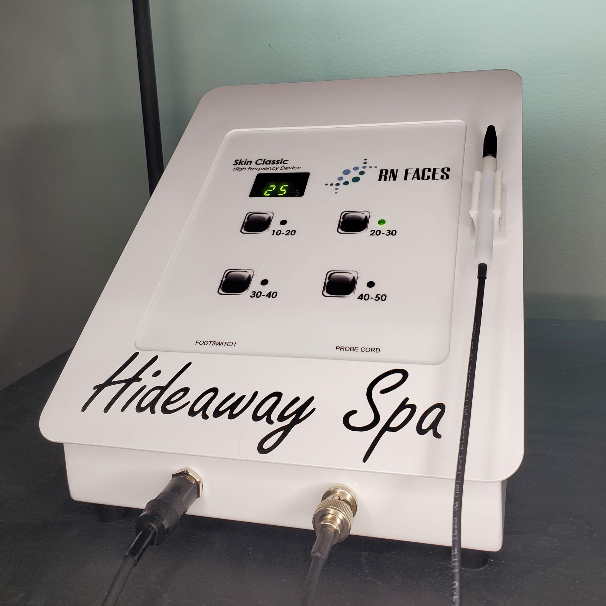
Our thermolysis treatment uses the Skin Classic device to deliver precise radiofrequency energy that instantly coagulates the blood vessels within the cherry angioma. Unlike methods that require days or weeks to see results, thermolysis provides immediate visible clearance — you watch the angioma lighten and flatten during the treatment itself.
How Our Treatment Works
During the procedure, a fine probe (sub-millimeter diameter) briefly contacts the cherry angioma for 1-5 seconds depending on size. The controlled radiofrequency energy instantly coagulates the dilated blood vessels, causing them to collapse. You'll see the bright red color visibly fade to pale pink or flesh-toned during treatment. Over the following 3-7 days, a tiny crust forms and naturally sheds as your body completes the healing process.
Instant Visible Results
Unlike lasers or cryotherapy that take days to weeks, thermolysis provides immediate gratification — the cherry angioma visibly lightens and flattens during the treatment itself. This instant feedback confirms successful treatment.10
Minimal Scarring
Thermolysis offers among the lowest scarring risks of all treatment methods. The precise, controlled energy delivery minimizes damage to surrounding tissue. Research shows excellent cosmetic outcomes with proper technique.10
Safe for All Skin Tones
Unlike certain lasers (PDL, KTP) that can cause pigmentation changes in darker skin, radiofrequency energy works safely on all skin types (Fitzpatrick I-VI) with equal effectiveness.
Highly Efficient for Multiple Angiomas
Treatment is extremely quick (1-5 seconds per angioma), making it ideal for addressing multiple cherry angiomas in a single session. Most people with 10-50 angiomas complete treatment in 10-20 minutes total.
Well-Tolerated Procedure
Most clients experience only a very brief pinch or sting lasting 105 seconds per angioma (only while the probe is on and in contact). The discomfort is momentary and well-tolerated without anesthesia, often described as a pin being pushed into your skin but not quite piercing. Much less painful than laser treatment purpura or cryotherapy blistering.
Usually One Session
Most cherry angiomas achieve complete clearance after a single treatment session with visible results immediately. Larger or more vascular angiomas may occasionally require a brief follow-up, but single-session clearance is the norm.
What to Expect During Your Visit
- Consultation & Assessment: We examine your cherry angiomas with a dermatoscope, confirm they're benign, and discuss your goals
- Treatment: Each angioma is treated individually with brief probe contact (1-5 seconds); you'll see it lighten immediately
- Immediate Aftercare: Treated areas may appear slightly pink with a tiny white coagulated spot immediately following treatment
- Natural Healing: Small crust forms and naturally sheds within 3-7 days; complete healing within 2 weeks
Session Duration: Most appointments last 10-30 minutes depending on the number of angiomas being treated. Because each one takes only seconds, even extensive treatment (20-50 angiomas) is completed quickly.
Aftercare Instructions
- Keep treated areas clean and dry for the first 24 hours
- Apply thin layer of antibiotic ointment (we provide bacitracin) as directed
- Avoid direct sun exposure during healing; use SPF 30+ after healing completes
- Critical: Do not pick, scratch, or forcibly remove tiny crusts — allow natural shedding to prevent scarring
- Avoid swimming, hot tubs, and excessive sweating for 48-72 hours
- Mineral makeup may be applied after 24 hours if needed
The Science Behind Thermolysis
Thermolysis uses radiofrequency energy (typically 13.56-27.12 MHz) to generate heat through ionic agitation within tissue fluids. This resistive heating causes targeted coagulation of blood vessels without cutting or vaporization.
For cherry angiomas specifically, the RF energy heats the hemoglobin in the dilated capillaries, causing them to coagulate (clot) and collapse instantly. The treated vessels cannot reform, so the angioma doesn't return in that exact spot. This is why you see immediate visible lightening during treatment — the blood is being coagulated in real-time.10
Learn more about thermolysis technology on our blemish correction page.
Frequently Asked Questions
Can you remove my cherry angiomas?
Yes, I can treat your cherry angiomas using thermolysis. Here's how it works: I use the Skin Classic device to deliver precise radiofrequency energy that coagulates the blood vessels within the angioma instantly. You'll see visible clearance during the treatment as the vessels collapse and the bright red color fades to pale pink or flesh tone (the red literally just pops away).
Your body then naturally completes the healing process over 3-7 days as a tiny crust forms and sheds. This is why thermolysis has such low scarring risk and provides instant visible results — there's no cutting, no waiting weeks to see if it worked, and the coagulated vessels cannot reform.
So technically, I perform the treatment that coagulates the vessels, and your body completes the healing process naturally. This approach produces clean results with minimal scarring and immediate gratification.
Do cherry angiomas grow back after treatment?
Successfully treated cherry angiomas do not regrow in the exact same spot because the coagulated vessels cannot reform. However, if you're prone to developing cherry angiomas due to age or genetics, new ones may appear in other areas over time.
Thermolysis does not prevent new cherry angioma formation — it addresses existing ones. The recurrence rate for thermolysis is quite low (5-10%) compared to other methods like cryotherapy (15-20%).9
Is thermolysis treatment painful for cherry angiomas?
Most people describe the sensation as a very brief pinch or sting lasting only 1-5 seconds per angioma while the probe is in contact, at this time it feels like a sharp pin being pressed to your skin. The discomfort is momentary and well-tolerated by most clients without any anesthesia.
Cherry angiomas typically require only seconds of treatment time (much faster than skin tags), making it one of the quickest and most comfortable removal methods available. Pain tolerance varies individually, but the brief contact time means even sensitive clients handle it well.
How many treatment sessions will I need?
The vast majority of cherry angiomas achieve complete clearance after a single treatment session. Unlike many other methods that require 2-3 visits, thermolysis typically provides instant visible results with one treatment.
Larger or more vascular cherry angiomas may occasionally require a brief follow-up treatment if the vessels are particularly deep or extensive, but single-session clearance is the norm.
Can I treat cherry angiomas at home?
Home treatment of cherry angiomas is strongly discouraged for several important reasons:
- Misdiagnosis risk: What looks like a cherry angioma might be amelanotic melanoma or another condition requiring medical care
- Bleeding: Cherry angiomas are vascular — improper treatment causes significant bleeding
- Infection: Non-sterile technique and equipment introduce bacteria
- Scarring: At-home methods almost always cause worse scarring than professional treatment
- Burns: "Cherry angioma removal pens" lack proper safety controls and often cause burns
Professional treatment ensures proper diagnosis, sterile technique, appropriate treatment parameters, and optimal cosmetic outcomes. The cost difference is minimal when you factor in the risks of home treatment.
Are cherry angiomas a sign of something serious?
Cherry angiomas themselves are entirely benign and have no relationship to cancer. They're an extremely common part of aging, with over 75% of adults over age 75 having them.1
However, sudden appearance of many cherry angiomas (called eruptive cherry angiomas) can sometimes indicate underlying conditions such as immunosuppression, graft-versus-host disease, multi-centric Castleman disease, or lymphoproliferative disorders.1
If you develop dozens of new cherry angiomas rapidly (over weeks/months rather than gradually over years), consult your family doctor for evaluation. For most people, cherry angiomas simply appear gradually as a normal part of aging with no underlying condition.
Can thermolysis be used on cherry angiomas anywhere on the body?
Yes. Thermolysis is safe and effective for cherry angiomas on all body areas including trunk (most common), arms, legs, neck, and face. The sub-millimeter probe tip allows precise treatment even in cosmetically sensitive areas.
Cherry angiomas rarely appear on hands, feet, or mucous membranes, but when they do, thermolysis can still treat them safely in most cases.
Learn more about how thermolysis works →
What's the difference between thermolysis and laser treatment for cherry angiomas?
Thermolysis uses radiofrequency energy to coagulate blood vessels, while lasers use focused light energy. Key differences include:
- Instant results: Thermolysis provides immediate visible clearance during treatment; lasers darken first, then fade over 1-2 weeks
- Cost: Thermolysis typically more affordable ($50-200 vs $200-500+ for laser)
- Skin tone safety: Thermolysis works on all skin tones; PDL and KTP lasers can cause pigmentation changes in darker skin (Nd:YAG safer but more expensive)2
- Pain level: Thermolysis brief sting (3-5 seconds); laser causes purpura (bruising) lasting 7-14 days
Both are effective methods. Pulsed dye laser is considered the gold standard by some dermatologists for minimal pain during treatment, but thermolysis offers comparable clearance rates with instant gratification, lower cost, and no post-treatment bruising.
Will my insurance cover cherry angioma removal?
Most insurance plans consider cherry angioma removal cosmetic and elective, so it's typically not covered unless the angioma is causing functional problems (e.g., vision obstruction, repeated traumatic bleeding interfering with daily life).
At Hideaway Spa, we offer transparent self-pay pricing with no insurance billing complexities. Our pricing structure makes professional treatment accessible and affordable for most budgets.
How long after treatment until I can exercise or swim?
Avoid swimming pools, hot tubs, saunas, and excessive sweating for 48-72 hours post-treatment to prevent infection and allow initial healing. Light exercise is fine immediately, but activities that cause heavy sweating should be postponed for 2-3 days.
After the small crust naturally sheds (typically 3-7 days), you may resume all normal activities. Apply sunscreen to treated areas during outdoor exercise once healed to prevent pigmentation changes.
Can I have multiple cherry angiomas treated in one visit?
Absolutely. In fact, thermolysis is particularly efficient for treating multiple cherry angiomas in a single session. Since each angioma only takes 3-10 seconds to treat, most clients with 10-50 angiomas complete their full treatment within 10-20 minutes.
We offer time-block pricing options (10 or 20 minutes) specifically for those addressing multiple angiomas, which is typically more cost-effective than per-lesion pricing.
What device do you use, and what devices are similar?
At Hideaway Spa, we use a Skin Classic device. Our main Blemish Correction page has detailed information about our machine and how it works.
Generally, the Skin Classic, Lamprobe, ThermoClear, SkinSheer, SIIT Pro, Perfect Touch, Skin Sheek, VascuTouch, Lamskin, and ThermoLo all use the same thermolysis/electrocautery principle to treat vascular lesions like cherry angiomas.
What is the healing process and aftercare like after a cherry angioma treatment?
Healing is typically quick for benign lesions: A small scab or crust usually forms within 24–72 hours and sheds naturally in 3–7 days, revealing fresh skin. Most visible healing (normal color and texture return) occurs within 7–21 days, though in some cases temporary pinkness or minor pigmentation changes may take several months to fully resolve.
We provide a detailed aftercare packet with your service, including these key instructions:
- Keep the area clean and dry initially; apply a thin layer of recommended antibiotic ointment to prevent infection.
- Avoid picking, scratching, or removing crusts prematurely — let them fall off naturally.
- Skip direct sun exposure, tanning, saunas, pools/hot tubs, heavy sweating, and makeup over crusted spots for 48–72 hours (or until healed).
- Use broad-spectrum SPF 30+ daily (even indoors) once the area has re-epithelialized — this is crucial to prevent prolonged or permanent pigmentation changes.
- Avoid harsh products, exfoliants, or irritants on the area during recovery.
Most clients see excellent cosmetic results with minimal visible signs after 1–2 weeks when following aftercare. If you notice signs of infection (increased redness, pus, swelling) or prolonged changes, contact us right away.
Is thermolysis safe during pregnancy or while breastfeeding?
Thermolysis is a superficial, localized treatment with no known systemic effects. It's generally considered safe for minor benign lesions when necessary, including during pregnancy (after the first trimester in some cases) and while breastfeeding, as there's no evidence of risk to the fetus or breast milk. However, due to limited specific studies and potential skin sensitivity changes during pregnancy, we recommend postponing non-essential treatments during active pregnancy as a precaution. For those planning pregnancy, there's no concern — it doesn't affect fertility or conception. Always disclose pregnancy, breastfeeding, or plans during consultation so we can assess and tailor accordingly (e.g., use smoke evacuation if needed). Safer alternatives like gentle skincare can be discussed if deferring.
What other types of blemishes can you treat?
Aside from cherry angiomas, we safely and effectively treat a variety of common benign skin concerns using our Skin Classic device. These include skin tags, sebaceous hyperplasia, milia, cholesterol deposits, age spots, and more. We also can treat a large selection of wrinkles, fine lines, scarring, texture or pigmentation issues with our Procell microchanneling device, a less traumatic form of microneedling using medical grade stem cell/peptide serums.
See all the Skin Classic's treatable skin conditions →
Learn about microchanneling for overall skin texture & rejuvenation →
About Erika — Certified Esthetician & Thermolysis Specialist
Erika Ziraldo brings over 15 years of experience in advanced skincare and has worked alongside dermatology teams using precision thermolysis, laser, and RF devices. She maintains specialized training in the Skin Classic system and takes an evidence-based, personalized approach to cherry angioma treatment in a private studio setting.
With hundreds of successful vascular lesion treatments completed, Erika combines technical expertise with a gentle, efficient approach. Her clients appreciate seeing immediate visible results during treatment rather than waiting weeks to see if the procedure worked. Learn more about Erika's background and credentials →
Informed Consent & Treatment Protocol
Informed written consent is required before all thermolysis treatments. During your consultation, we'll review the procedure, expected outcomes, potential side effects, and aftercare requirements to ensure you have complete understanding before proceeding.
We request permission for before/after photography for clinical records and potential marketing use; clients may decline public sharing while maintaining private clinical documentation.
Medical Referral Protocol
Any growth exhibiting irregular vascular patterns, asymmetry, rapid growth, spontaneous bleeding, or other clinically suspicious features will be referred for dermatological evaluation before treatment. Thermolysis is performed exclusively on confirmed benign cherry angiomas within cosmetic scope of practice.
Disclaimer: Hideaway Spa provides cosmetic thermolysis for benign vascular lesions confirmed through dermatoscopic assessment. This is not a medical service and does not replace physician evaluation. When medical assessment is indicated, we provide referral recommendations and defer treatment pending clearance. Erika Ziraldo is a certified esthetician, not a medical doctor.
